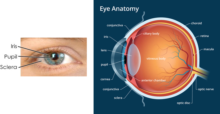
Eye Anatomy
The retina of the eye is a thin layers of diverse nerve cells inside the eye, which is like a film compared to the camera. When you see an object or text, the image is transmitted to the brain by the retina. The retina converts light energy entering our eyes into electrical energy and transmits it to the brain through optic nerves. The retina is made up of over 100 million light-sensing cells (photoreceptor cells), over a million optic nerve cells, and numerous cells that serve as wires connecting them, making it one of the most sophisticated tissues in our body. About 30% of brain cells are used to process visual information in the retina.
Retina structure
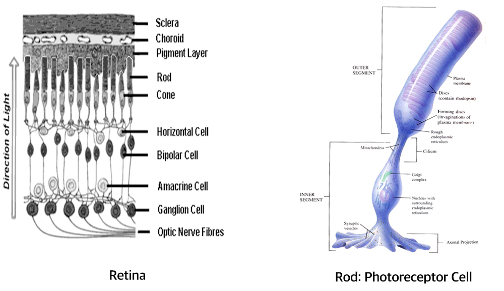
The retina is made up of eight different cells including rod, cone, retinal pigment epithelium, ganglion, amacrine, horizonthal, Müller, and bipolar cells. Light is sensed in rods and cones, and once they die, they are not regenerated.
Eyes and disease
Representative retinal diseases include macular degeneration, diabetic retinopathy, retinitis pigmentosa, and retinal detachment.
Age-related macular degeneration is the most common cause of blindness in people over the age of 50s, with an estimated 15 million cases in the United States alone.
Diabetic retinopathy, a complication in diabetic patients, is the most common cause of blindness in the population aged 20 to 74 in the United States.
Retinitis pigmentosa is a typical hereditary retinal disease, and although the cause is gradually being elucidated, it is still one of the most difficult diseases to treat.
Retinal detachment is a disease in which the retina separates from the retinal pigment epithelium, and in severe cases, blindness and geographic atrophy may occur. Because the retina is located deep inside the eye, it is difficult to determine whether there is an abnormality only with a general external examination.
For the examination and treatment of retinal diseases, special equipment and the help of specialists are required.
COPYRIGHT ⓒ JULIA LABORATORY. ALL RIGHTS RESERVED. DESIGNED BY MARVEL WORKS
 ENG
ENG  KOR
KOR